


Cardiapoda placenta
Roger R. Seapy
Introduction
Cardiapoda placenta is the larger of the two species of Cardiapoda, achieving a maximal size of about 110 mm. Features that distinguish C. placenta from C. richardi include: numerous gills (>20) that form a median crest on the visceral nucleus, fin sucker in both males and females, a tail that terminates in 12 finger-like extensions that can be expanded and contracted, and a narrowly triangular eye shape.
Brief Diagnosis
A species of Cardiapoda with:
- Gills numerous (>20), forming a median crest on the visceral nucleus
- Fin sucker present in both sexes
- Tail terminates in 12 finger-like contractile extensions
- Shape of eye, viewed dorsally, narrowly triangular
Characteristics
- Body morphology
- Gills numerous (>20), forming an arc or crest on the posterior and dorsal surface of the visceral nucleus
- Fin sucker present on both males and females
- Tail terminates in 12 fan-shaped, reddish-brown finger-like extensions that are normally expanded, but can contract when the animal is disturbed
Click on an image to view larger version & data in a new window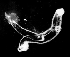
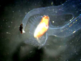
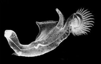
Figure. Left: black and white, in-situ photograph of Cardiapoda placenta, left side of body; note the fan-shaped, ventral tail extensions. © R. Gilmer. Right: Photograph of posterior portion of C. placenta, left side of body; note that the tail extensions are retracted. © A. Connell
- Gills numerous (>20), forming an arc or crest on the posterior and dorsal surface of the visceral nucleus
- Radula with a tricuspid central (rachidian) tooth. The cusps are pointed, comparable in length, and the lateral cusps angle outward somewhat from the median cusp. This tooth shape differs from that in C. richardi in that the lateral cusps are shorter than the median cusp and the lateral cusps do not angle outward.
Click on an image to view larger version & data in a new windowClick on an image to view larger version & data in a new window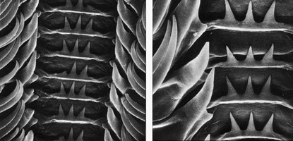
Figure. Radula of Cardiapoda placenta. Left: Lower magnification view; proceeding inward in each tooth row, the photograph shows the terminal parts of the elongate paired marginal teeth, the elongate lateral tooth, and the central rachidian tooth. Right: high magnification view of three central rachidian teeth. Photograph from Thiriot-Quiévreux (1975, Fig. 5A,B) © 1975 C. Thiriot
- Shell
- Adult shell microscopic; following metamorphosis teleoconch is formed at right angles from aperture of protoconch (larval shell)
Click on an image to view larger version & data in a new window
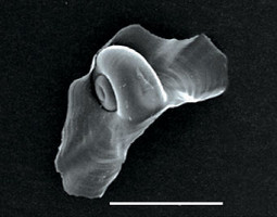
Figure. Adult shell of Cardiapoda placenta. Left: sketches of shell, viewed from the right side and aperture, respectively. Right: Scanning electron micrograph of shell, viewed from frontal. Note in both images the large size of the teleoconch in contrast with the early adult shell used on the Cardiapoda page (drawn by Tesch, 1949). Shell sketches © 1976 S. van der Spoel; SEM photograph © Orso Angulo
- In adult animals, the shell is located at the posterior end of the visceral nucleus, beneath (ventral to) the most posterior gills.
Click on an image to view larger version & data in a new window
Figure. Posterior portion of the stalked visceral nucleus in Cardiapoda placenta. The posterior portion of the dorsal gills and the postero-ventral location of the imbedded shell are indicated in the photograph. Photograph modified from Thiriot-Quiévreux (1975, Fig. 4B) © 1975 C. Thiriot
- Adult shell microscopic; following metamorphosis teleoconch is formed at right angles from aperture of protoconch (larval shell)
- Larva
- Larval body light brown to clear; digestive gland yellow; velum bordered by brown pigment, with two large brown patches on each lobe (Thiriot-Quiévreux, 1975)
- Larval shell globular, with a large oval aperture (below left). The spire has two spiral ridges on the second whorl (below right). In C. placenta the outermost of these ridges is shorter than that in C. richardi (see larval shell on C. richardi page).
Click on an image to view larger version & data in a new window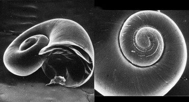
Click on an image to view larger version & data in a new windowFigure. Larval shell of Cardiapoda placenta viewed from the right side, tilted (left) and spire (right). Photographs modified from Thiriot-Quiévreux (1975, Fig. 1F,G) © 1975 C. Thiriot
Comments
In-situ field observations of C. placenta by Ronald Gilmer (reported in Lalli and Gilmer, 1989) revealed that both sexes are brightly colored with reddish-brown spots covering the body, although the number and brightness of these spots are greater in females. The pigment spots were seen to be highly contractile, changing rapidly from 0.5 to 2 mm in diameter. Also, the terminal finger-like expansions on the tail varied from reddish-brown to black, and flashed during prey capture.
The feeding behavior of C. placenta was observed on 38 occasions by SCUBA divers in the Sargasso Sea and Florida Current (Hamner, et al., 1975). The most frequently encountered prey were salps. Attack of a prey item was seen once in the field and twice in aquaria (using hand-collected animals). In each case the prey's silhouette appeared to have been located visually from beneath by the upward-searching predators from as far away as 60 cm. In each case, the predator swam rapidly (up to 40 cm/sec) up to the prey and captured it with the buccal cones, followed by ingestion (taking about 10 min) using the radula and peristaltic movements of the proboscis.
References
Hamner, W. M., L. P. Madin, A. L. Alldredge, R. W. Gilmer, and P. P. Hamner. 1975. Underwater observations of gelatinous zooplankton: Sampling problems, feeding biology, and behavior. Limnology and Oceanography 20: 907-916.
Lalli, C. M. and R. W. Gilmer. 1989. Pelagic snails. The biology of holoplanktonic gastropod snails. Stanford University Press, Stanford. 259 pp.
Spoel, S. van der. 1976. Pseudothecosomata, Gymnosomata and Heteropoda (Gastropoda). Bohn, Scheltema and Holkema, Utrecht. 484 pp.
Thiriot-Quiévreux, C. 1975. Observations sur les larves et les adultes de Carinariidae (Mollusca: Heteropoda) de l'Océan Atlantique Nord. Marine Biology 32: 379-388.
Title Illustrations

| Scientific Name | Cardiapoda placenta |
|---|---|
| Location | Florida Current |
| Specimen Condition | Live Specimen |
| Sex | Female |
| Life Cycle Stage | adult |
| View | right side |
| Size | 65 mm |
| Copyright | © Ronald Gilmer |
About This Page

California State University, Fullerton, California, USA
Correspondence regarding this page should be directed to Roger R. Seapy at
Page copyright © 2008
 Page: Tree of Life
Cardiapoda placenta .
Authored by
Roger R. Seapy.
The TEXT of this page is licensed under the
Creative Commons Attribution License - Version 3.0. Note that images and other media
featured on this page are each governed by their own license, and they may or may not be available
for reuse. Click on an image or a media link to access the media data window, which provides the
relevant licensing information. For the general terms and conditions of ToL material reuse and
redistribution, please see the Tree of Life Copyright
Policies.
Page: Tree of Life
Cardiapoda placenta .
Authored by
Roger R. Seapy.
The TEXT of this page is licensed under the
Creative Commons Attribution License - Version 3.0. Note that images and other media
featured on this page are each governed by their own license, and they may or may not be available
for reuse. Click on an image or a media link to access the media data window, which provides the
relevant licensing information. For the general terms and conditions of ToL material reuse and
redistribution, please see the Tree of Life Copyright
Policies.
- First online 12 February 2008
- Content changed 08 July 2008
Citing this page:
Seapy, Roger R. 2008. Cardiapoda placenta . Version 08 July 2008. http://tolweb.org/Cardiapoda_placenta/28744/2008.07.08 in The Tree of Life Web Project, http://tolweb.org/


