


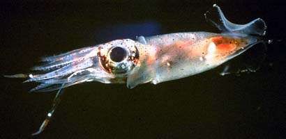
Pterygioteuthis microlampas
Annie Lindgren, Richard E. Young, and Katharina M. Mangold (1922-2003)
Introduction
Pterygioteuthis microlampas and P. gemmata are very similar but are most easily separated by the smaller size of adult P. microlampas and fewer number of hooks on the arms of males of this species (Riddell, 1985).
A Pterygioteuthis with ...
- hooks in 1 series (ventral row) on arms I-III .
- arms III with 2-3 hooks in males.
- 4 tentacular photophores.
Characteristics
- Arms - hooks on arms I-III in 1 series (ventral). Sheath (fleshy covering) is greatly enlarged on all hooks in males.
- Arms I
- 3-6 pairs of suckers proximal to hooks.
- Hooks: 1 in males, 3 in females.
- Small, normal (non-globular) suckers distal to hooks.
Click on an image to view larger version & data in a new window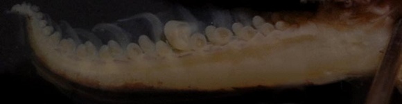
Figure. Side view of left arm I from a male P. microlampas. Note single enlarged hook sheath. ©
- Arms II
- 5 pairs suckers proximal to hooks.
- Hooks: 1-2 in males, 3-5 in females
- 4-8 pairs small suckers distal to hooks.
Click on an image to view larger version & data in a new window
Figure. Oral view of a right arm II for a male P. microlampas. Note enlarged ventral hook sheaths. © Annie Lindgren
- Arms III
- Suckers proximal to hooks: 3 pair in males, 4-5 pair in females.
- Hooks: 2-3 in males, 3-5 in females.
- 4-7 pairs small suckers distal to hooks.
Click on an image to view larger version & data in a new window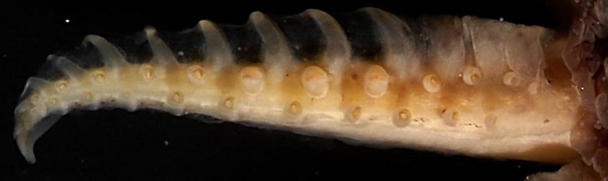
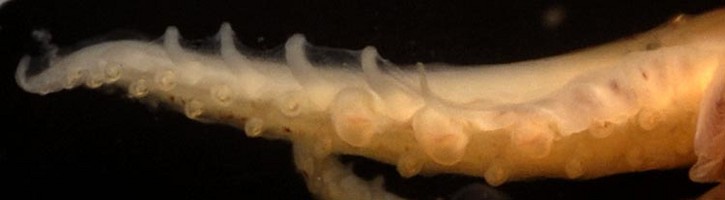
Figure. Oral view of left arms III for P. microlampas. Top - Female with 3 hooks. Bottom - Male with 3 hooks and enlarged sheaths. ©
- Arms IV (click here for images).
- 2 series small suckers in males and females.
- Left arm IV hectocotylized. Hectocotylus plate with many small teeth.
- 2 series small suckers in males and females.
- Arms I
- Tentacles
- Suckers in 4 series, but proximal 2-4 rows have enlarged dorsal suckers (click here for image).
- Suckers in 4 series, but proximal 2-4 rows have enlarged dorsal suckers (click here for image).
- Head
- Buccal crown with papillae (few to no ridges - click here for images).
- Photophores
- Eye with 10 large (9 visible on left eye of photograph) and 4 small photophores (visible on right eye of photograph - click here for images).
- Tentacles with 4 embedded photophores (1 large, spherical photophore at bend in base of tentacular stalk; 2 small spherical photophores along stalk, 1 small spherical photophore at club base - click here for images).
- Eye with 10 large (9 visible on left eye of photograph) and 4 small photophores (visible on right eye of photograph - click here for images).
- Pigmentation
- Tentacular chromatophores are concentrated at the base (or bulge) of tentacle stalk, continue up along the stalk in a single row, concentrated again at the tentacle club base, and cover the aboral surface of the tentacular club.
Click on an image to view larger version & data in a new window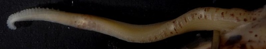
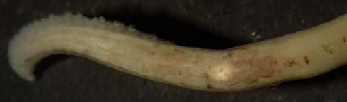
Figure. Top - Side view of a tentacular chromatophores of P. microlampas. Bottom - Close-up view of aboral tentacle club chromatophores. ©
- Funnel chromatophores begin dorsolateral to locking apparatus, and continue up and across the ventral funnel surface.
Click on an image to view larger version & data in a new window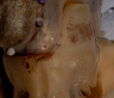
Figure. Ventral view of a male P. microlampas. Note large chormatophores on ventral surface. ©
- Size
- Females mature at 18-20 mm ML; males at 17 mm ML (Riddell, 1985).
- Maximum length about 23 mm ML.
* Characters shared with P. gemmata.
Comments
Life history
Paralarvae
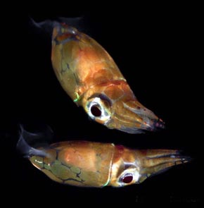
Figure. Paralarval P. microlampas. Top -Ventral view, 2.9 mm ML. Drawing modified from Young, et al., 1992. Bottom - Anterodorsal, lateral views. In situ photographs by Jeffrey Milisen.
Young paralarvae are most easily recognized by:
- Large, dark chromatophore at the base of the tentacular club.
- Small size of the ocular photophores.
- Pair of chromatophores of the ventral surface of the funnel (present only in advanced paralarvae; colored grey in the illustration as it is hidden beneath the mantle).
- Numerous chromatophores on the mantle.
Although paralarvae are easily damaged in plankton nets, character number 1 is usually apparent. Compare with paralarvae of Pterygioteuthis giardi and Pyroteuthis addolux.
Juveniles
Juveniles of GL< 10 mm have several adult characteristics:
- Buccal membrane with papillae
- 2-3 hooks on arms III
- Founder chromatophores on funnel (see picture below)
Click on an image to view larger version & data in a new window
Figure. Ventral surface of a juvenile P. microlampas. Note 4 founder chromatophores: 1 just anterior to locking apparatus, and 2 on ventral funnel surface. ©
Distribution
Vertical distribution
In Hawaiian waters P. microlampas is found mostly between depths of 450-500m during the day and 50-100 m at night (Young, 1978).
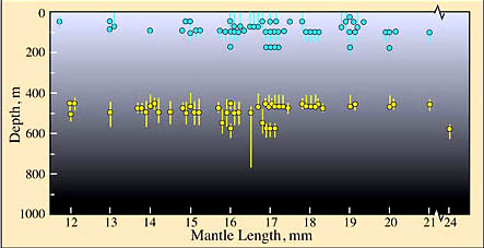
Figure. Vertical distirbution of P. microlampas, Hawaiian waters. Yellow dots - Modal depth of trawl, day capture. Blue dots - Modal depth of trawl, night capture. Bars - Depth range of trawl. Chart modified from Young, 1978.
Geographical distribution
This species was first described from Hawaiian waters (Berry, 1913) and has since been reported from waters near New Zealand north of the tropical convergence (ca. 28° S. Lat.). At this locality it barely overlaps with the more southernly P. gemmata as seen in the data from Riddell (1985).
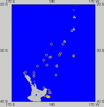
Figure. Geographical distribution of Pterygioteuthis spp. near New Zealand. Yellow circles represent captures of P. gemmata and white triangles captures of P. microlampas. Chart modified from Riddell, 1985.
The world map shows some of the general localities (white circles) where P. microlampas has been captured. Localities where pyroteuthids, other than this species, have been captured are represented by yellow crosses. Only one record per general locality is included (records listed here). P. microlampas presumably occurs throughout much of the tropical Pacific but seems to be absent from the Eastern Tropical Pacific. No regional differences in taxonomic characteristics are known.
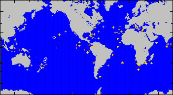
Figure. Geographical distribution of P. microlampas (white circles).
References
Berry, S. S. 1913. Diagnoses of new cephalopods from the Hawaiian Islands. Proc. U. S. Nat. Mus. 37: 407-419.
Lindgren, A.R. 2010. Systematics and distribution of the squid genus Pterygioteuthis (Cephalopoda: Oegopsida) in the eastern tropical Pacific Ocean. Journal of Molluscan Studies, 76(4): 398-398.
Riddell, D. J. 1985. Enoploteuthidae of the New Zealand Region. Fisheries Research Bulletin. New Zealand Ministry of Agriculture and Fisheries, No.27: 1-52.
Young, R. E., K. M. Mangold and M. Vecchione. 1992. The enoploteuthid group of families. P. 55-66. In: Sweeney, M. J., C. F. E. Roper, K. M. Mangold, M. R. Clarke and S. V. Boletzky (Eds.). "Larval" and juvenile cephalopods: A manual for their identification. Smithson. Contr. Zool. No. 513.
Title Illustrations

| Scientific Name | Pterygioteuthis microlampas |
|---|---|
| Location | Hawaiian waters |
| View | ventrolateral |
| Image Use |
 This media file is licensed under the Creative Commons Attribution-NonCommercial License - Version 3.0. This media file is licensed under the Creative Commons Attribution-NonCommercial License - Version 3.0.
|
| Copyright |
© 1996

|
About This Page
Ohio State University, Columbus, Ohio, USA
University of Hawaii, Honolulu, HI, USA
Katharina M. Mangold (1922-2003)
Laboratoire Arago, Banyuls-Sur-Mer, France
Correspondence regarding this page should be directed to Annie Lindgren at and Richard E. Young at
Page copyright © 2016 , , and Katharina M. Mangold (1922-2003)
All Rights Reserved.
- Content changed 16 November 2016
Citing this page:
Lindgren, Annie, Richard E. Young, and Katharina M. Mangold (1922-2003). 2016. Pterygioteuthis microlampas . Version 16 November 2016 (under construction). http://tolweb.org/Pterygioteuthis_microlampas/19752/2016.11.16 in The Tree of Life Web Project, http://tolweb.org/


