- Photophores
- Eyeball: lateral series= 8 separate organs (may coalesce to irregular stripe + 1); intermediate series = stripe with nearly separate photophore at posterior end; medial series = stripe.
- Viscera: none
 Click on an image to view larger version & data in a new window
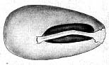
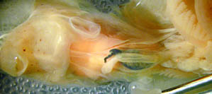
Figure. Left - Ventral view of the digestive gland, ink sac and intestine of C. mega showing absence of photophores on the ink sac. Drawing from Voss (1967). Right - Photograph of the same region of C. mega. Photograph by R. Young.
- Club-tip photophore large (exposed photogenetic surface much more than half the photophore length), with little or no terminal papilla. Click on an image to view larger version & data in a new window
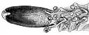
Figure. Oral view of the club-tip photophore of C. mega. Drawing from Voss (1967).
- Distal most cushion-like photophore of stalk partially embedded, located just proximal to first club sucker; no photophores embedded in aboral surface of club.
- Pigmentation
- Club-tip photophore with heavy epidermal pigmentation in covering lids and adjacent tissue.
- All other club pigment in functional chromatophores.
- Club sucker stalks without pigment.
- Buccal membrane pigmented (epithelial pigmentation on oral and aboral surfaces).
- Olfactory organ unpigmented in small subadults but with pigment in chromatophores at 115 mm ML.
- Arms
- Arms I 30-50% of ML.
- Arms III 40-60% of ML.
- Arms IV 120-140% of ML.
- Large arm suckers with 20-30 teeth around entire ring. Teeth slender, pointed, separate distally; truncated, separate laterally; broadly rounded, in contact proximally.
- Largest suckers not globular.
Scanning electron micrographs of the arm suckers can be seen here.
- Tentacular clubs
- Club length 84% of ML.
- Suckers with 15 pointed teeth over distal 3/4 of ring; enlarged, conspicuous central tooth; teeth just lateral to central tooth smallest of distal teeth; club suckers equal to or slightly larger than largest arm III sucker.
- Stalks weakly divided into two parts: proximal portion with thick, delicate keel with terminal swelling (=flag). Stalks of lateral suckers about twice as long as stalks of medial suckers.
- Protective membranes
- Membranes in two sets; proximal set slightly larger than distal set.
- Proximal set short (5-7% of club length), with 12-15 trabeculae, numbers 5-11 adjacent (in contact) and more elongate than the others. At broadest point, four trabeculae present between adjacent lateral suckers.
- Distal set long (93-95% of club length) with 58-70 broad, well-separated trabeculae. Proximal 1 or 2 trabeculae bifurcate at the tip. Trabeculae arise alternately with lateral suckers; shaped like broad, truncated bands that narrow slightly distally; separated by width of about one trabecula.
Scanning electron micrographs of the club suckers can be seen here.
Click on an image to view larger version & data in a new window
Figure. Oral view of medial (left) and lateral (right) club suckers and stalks of C. mega. Drawing from Voss (1967)
Click on an image to view larger version & data in a new window
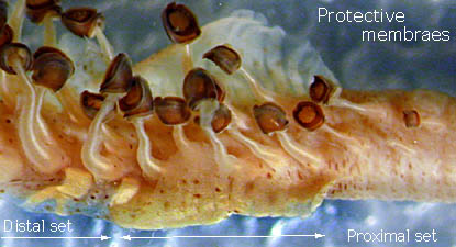
Figure. Oral views of the tentacular club of C. mega. Top - Drawing from Voss (1967). Bottom - Photograph of short proximal segment by R. Young.
- Head
- Head length 28-33% of ML
- Funnel
- Tragus of the funnel-locking apparatus well developed but not as prominent as in Chiroteuthis sp. B, with broad base. Click on an image to view larger version & data in a new window

Figure. Frontal view of the funnel locking-apparatus of C. mega. Drawing from Voss (1967)
- Tragus of the funnel-locking apparatus well developed but not as prominent as in Chiroteuthis sp. B, with broad base.
- Fins
- Fin length ca. 45% of ML
- Fin width ca. 35% of ML
Comments
Fins with distinct, small posterior lobes; marginal membrane of tail thick, lacks trabeculae. The penis on mature males extends well beyond the mantle opening.


