Two hooks of Kondakovia longimana were examined with the scanning electron microscope (SEM). Their relative sizes are seen in the phtotgraph below. The specimen available had lost some of the club hooks so we were unable to select hooks comparable to those of other genera. We examined the 11th hook from the proximal end of the club in the ventral series (i.e., hook V11) but we estimate that the largest hook on the club was probably at position 7 or 8. We examined one hook from the dorsal series (hook D5). In addition, the hooks had been damaged by fixation as can be seen in the lumpy claw tip of hook V11.

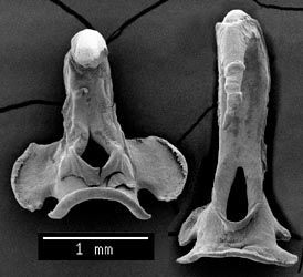
Figure. Frontal views of hooks D5 (left) and V11 (right) of K. longmana, 195 mm ML, ELTANIN sta. 1392, NMNH specimen. SEM photograph by R. Young.
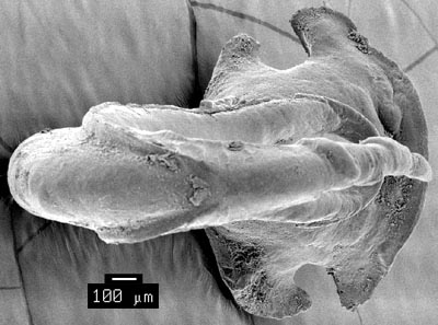
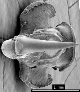
Figure. Top-frontal views of the two hooks (V11, left; D5, right). The lateral "grooves" of hook V11 meet in the midline and the top wall of the groove is absent. The claw beyond the grooves is relatively long. Note the strong constriction of the claw in D5 here and in the photograph below. This constriction is probably a fixation artifact. However, in D5 the converging lateral grooves can still be seen as thin lines forming a V-shape. In V11 the proximal lip lacks a "hair lip" and the lateral lobes are well developed and slightly different in size. SEM photographs by R. Young.
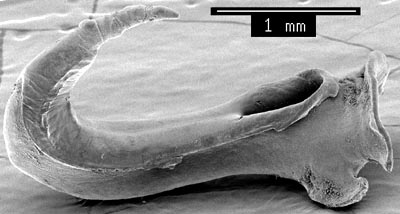
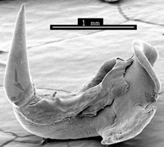
Figure. Side views of the two hooks (V11, left; D5, right). The constriction near the base of the claw of D5 is more apparent in this view. See comments above. SEM photographs by R. Young.
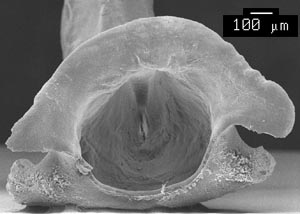
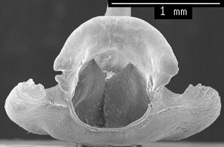
Figure. Attachment-site views of the two hooks (V11, left; D5, right). SEM photograph by R. Young.
Comments
The joining of the claw grooves is seen only in this squid and Notonykia africanae. The hooks are distinctive in having a long terminal region of the claw without lateral grooves and are similar to most species of Onykia examined in the near symmetry of the lateral lobes and the absence of a hair-lip on the proximal lip of the ventral-series hook.


