
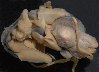
Figure. Side view of the second specimen of Iridoteuthis sp. A, male, damaged, mantle everted. Photograph by R. Young.
- Arms
- Arms IV tips in both sexes with numerous small suckers which seem in 2 series in female and multiple series in male (see photograph below).
- Deep web between all arms; deepest between arms I and least deep between arms IV.
- Web in sector A extends along arms nearly to tip of each arm I; web in sector B not as extensive; web in sector C extends well along arm III (but not arm II); web sector D extends along arm IV as a lateral membrane that remains broad to arm tip; web in sector E short. The webs are not in perfect condition and are difficult to interpret, therefore, possible differences between sexes were not apparent.
- Arms III with a thin, broad, muscular keel (flag) that remains broad to arm tips; arms I and II without distinct keels or flags.
- Bases of all arms and webs, proximal to first sucker, in females only, are covered with numerous small papillae (see photograph below).
- Arm suckers globular with smooth inner rings (see photograph below).
- Arms, other than arms I in males, with delicate, protective membranes, often discontinuous, sometimes forming flaps.
Click on an image to view larger version & data in a new window
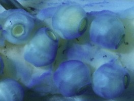
Figure. Left - Oral view of the arm IV tip of Iridoteuthis sp. A, male, showing numerous small suckers in several series. Note large lateral membrane. Right - Oral view of large arm suckers of left arm I of Iridoteuthis sp. A, male, stained with methylene blue stain. Photographs by R. Young.
More pictures of the arms can be seen here.Click on an image to view larger version & data in a new window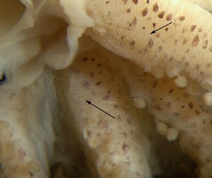
Figure. Oral view of the papillate region at the bases of the arms in female Iridoteuthis sp. A. Arrows point to the papillate bases of arm III (top) and arm I (bottom). Photograph by R. Young.
- Tentacles
- Tentacular clubs with small suckers in many irregular series; apparently present proximally along ventral margin, spreading distally over oral surface.
- Club suckers of unequal size with proximal suckers larger than distal suckers; some transverse variation occurs in distal half of club with those of ventrally larger than dorsally.
- Tentacular organ begins proximal to club and extends nearly to curled club tip.
Click on an image to view larger version & data in a new window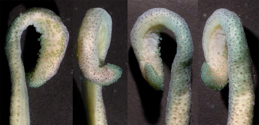
Figure. Various views of the tentacular club of Iridoteuthis sp. A, female. This series shows the unflattened club rotated into various positions. The club was lightly stained with methylene blue stain. Photograph by R. Young.
Click on an image to view larger version & data in a new window
Figure. Various views of the tentacular club of Iridoteuthis sp. A, female. The coiled club was flattened with a glass slide which distorts the club shape. Left arrow points to tentacular organ, two other arrows point to the beginning and end of the tentacular organ. Photograph by R. Young.
Click on an image to view larger version & data in a new window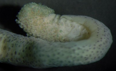
Figure. Oral view of the distal region of the club of Iridoteuthis sp. A, showing the termination of the tentacle organ very near the recurved club tip. Photograph by R. Young.
- Head
- Eyes very large and with secondary eyelid.
- Olfactory organ bulbous, with chromatophores on head, lies in a circular pocket in male; could not be detected in female and found only on the right side in male.
- Head with muscular lateral funnel adductor (see photograph below).
Click on an image to view larger version & data in a new window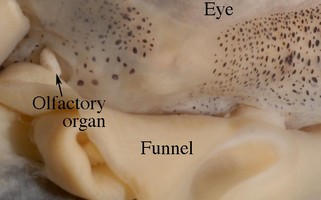
Figure. Side view of the posterolateral corner of the head of Iridoteuthis sp. A, mature male. Photograph by R. Young.
- Eyes very large and with secondary eyelid.
- Funnel
- Funnel valve present.
- Funnel component of funnel/mantle locking-apparatus with broad groove and deep pit near anterior end of groove; mantle component with high, angled ridge at anterior end, matching deep pit of funnel component.
Click on an image to view larger version & data in a new window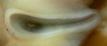
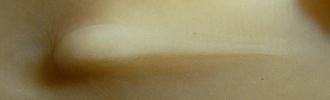
Figure. Funnel/mantle locking-apparatus of Iridoteuthis sp. A, female. Top - Frontal view of funnel component. Middle - Frontal view of mantle component. Bottom - Side view of mantle component. Photographs by R. Young.
- Mantle
- Mantle broadly fused to head dorsally; fusion reaches to posterior edge of eyes at level of lateral midpoint of each eye.
- Dorsal mantle with distinct hump (see left title photograph).
- Ventral mantle shield large, about 80% of ML.
- Mantle broadly fused to head dorsally; fusion reaches to posterior edge of eyes at level of lateral midpoint of each eye.
- Fins
- Posterior margin of fin with sharp corner. Click on an image to view larger version & data in a new window
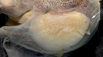
Figure. Dorsal view of the left fin of Iridoteuthis sp. A, female, showing sharp posterior angle. Photograph by R. Young.
- Posterior margin of fin with sharp corner.
- Photophores
- Large visceral photophore present.
- Photophore papillae each consist of two large tubes located at lateral edges of photophore; do not penetrate photophore "lens" apparently entering glandular photophore between "lens" and ink sac.
Click on an image to view larger version & data in a new window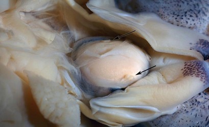
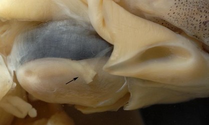
Figure. Visceral photophore pores and tubes (arrows) of Iridoteuthis sp. A. Top - Ventrolateral view, female. Bottom - Lateral view, male. Photographs by R. Young.
- Large visceral photophore present.
- Pigmentation
- Unpigmented strip on posteroventral surface of head from funnel to edge of secondary eyelid (see photograph below).
- Anteroventral surface of funnel with chromatophores; chromatophores absent from posteroventral, lateral and dorsal surfaces of funnel (see photograph below).
- Outer mantle chromatophores extextend slightly into mantle cavity along ventral mantle edge (see photograph below).
- Silver band on mantle approximately horizontal.
- Scattered chrmatophores on dorsal surface of fins except longitudinal strip near base (see photograph above of fin); none on ventral surfaces.
- Chromatophores present on all surfaces of arms.
- Distal half of aboral surface of tentacle with numerous chromatophores. Numbers diminish in proximal half, disappearing near tentacle base (see photograph below).
Click on an image to view larger version & data in a new window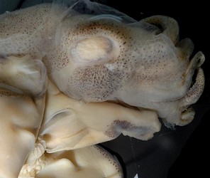
Figure. Side view of head, funnel and inner edge on mantle margin of Iridoteuthis sp. A, male, showing chromatophore patterns. Note loose cuticle on head and mantle. Photograph by R. Young.
Click on an image to view larger version & data in a new window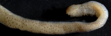
Figure. Aboral view of the distal region of the tentacle of Iridoteuthis sp. A, female, showing chromatophore pattern. Photograph by R. Young.
- Measurements, mm
Arm lengths measured from proximal-most sucker.Palinstru 03/10/2 Palinstru 03/10/1
Sex Male
Female
Dorsal mantle length* 23 (estimate) 23 Ventral mantle length ND 23 Lateral mantle length ND 15.5 Eye diameter ND
~9 Fin length ND
14 Fin width ND
10.5 Arm I length** 13 9 Arm II length
13 10 Arm III length
13 9 Arm IV length
9 10 Club length
5 4 Diam. largest suckers,
arms I-III0.7 0.4
Diam. largest suckers,
arms IV
0.4 0.3 Sucker counts: Arms I 30 ND
Web depth: Sector A 5 ND
Sector B
5 ND
Sector C
4 ND
Sector D
5 ND
Sector E
2.5 ND
ND - No data


