The patterns formed by alveolae and the thecal plates sometimes contained in them (tabulation) have been used in the taxonomy of dinoflagellate motile stages for more than 100 years. Six fundamental types of tabulation can be recognized (Figs A-E):
 |  |  |  | 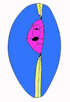 |
| Fig. A. Gymnodinoid tabulation | Fig. B. Suessoid tabulation | Fig. C. Peridinoid / gonyaulacoid tabulation | Fig. D. Dinophysoid tabulation | Fig. E. Prorocentroid tabulation. Periflagellar area with tiny platelets (in pink) |
 Click on an image to view larger version & data in a new window | Click on an image to view larger version & data in a new window | |||
| Drawings modified after Fensome et al. 1993. © Mona Hoppenrath | ||||
- Gymnodinoid. Alveolae are numerous and often hexagonal, the girdle and sulcus being the only clearly distinguishable series. Thecal plates are entirely absent.
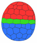
- Suessioid. Alveolae are arranged in 6-11 latitudinal series. The number of plates per series, or even the number of series, varies with species. The cingulum is well marked, and it may contain one or two rows of plates. Thecal plates are delicate.
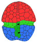
- Peridinioid and Gonyaulacoid. Alveolae are arranged in five distinct primary latitudinal series. They contain true thecal plates, termed from apex/anterior to antapex/posterior: apicals, precingulars, cingulars (girdle), postcingulars, and antapicals. Plates lying between these series are termed intercalaries (anterior or posterior on the epi- or hypotheca respectively), and those lying within the sulcus are sulcals. The midventral epithecal plate often spans both the precingular and apical series. By convention it has been termed the first apical plate. At the apex an apical pore complex (APC) is often present, consisting of an outer (Po) and inner (Pi) pore plate, and a small pre-apical platelet (Pp, canal plate) is often present in peridinioids. Apical plates are those that contact the APC. Peridinioid tabulations are defined by a more-or-less symmetrical first apical plate and by the presence of two antapical plates, in gonyaulacoid tabulations the first apical plate is asymmetrical and there are 2-4 fundital plates.
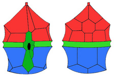
- Dinophysoid. The theca is fundamentally divisible into two halves by a vertical sagittal suture, but a girdle and sulcus are “superimposed” on it, separating an epitheca and hypotheca. There are small plates on the ventral surface of the epitheca, hypotheca, and in the sulcus around the single large flagellar pore. A simple apical pore is located on the ventral side of the epitheca. The arrangement of the plates varies little within the group. Lists (ridges or extensions of the edge of thecal plates) along the girdle and sulcus edges may be prominent and developed to an extraordinary degree in some genera, producing very bizarre forms. Some genera (e.g. Ornithocercus) form a “phaeosome chamber” with the girdle lists that contains extracellular coccoid cyanobacteria.
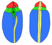
- Prorocentroid. The theca is composed of two large plates, the valves, which join along a toothed margin, the sagittal suture. A cluster of tiny plates (periflagellar platelets) in a regular arrangement surrounds the two pores. The desmokont flagella arise from one pore. The periflagellar platelets lie principally in an excavation of the right valve. A spine/tooth or protrusions/wings can arise from periflagellar platelets.
A convention, the “Kofoid System” of tabulation, is currently in universal use (Fig. F). The plates in each latitudinal series are numbered from the cell’s left to right, beginning with the plate closest to the midventral position. It also uses a notation to designate the series, using primes to indicate the apical ('), precingular (''), postcingular ('''), and antapical ('''') plates, both when labelling plates on figures and when producing a plate formula. The latter is a listing of the total number of plates in each series for a species or genus. Thus Peridinium is represented by APC, 4', 3a, 7'', 5C + t, 6S, 5''', 2'''' and Gonyaulax by APC 3', 2a, 6'', 6C + t, 6S, 6''', 1p, 1''''. Cingulars (C), sulcals (S), anterior intercalaries (a), and posterior intercalaries (p) are designated by letters. The t plate is a small transitional plate between the cingulars and the sulcals at the proximal end of the girdle in peridinioids and at the distal end in gonyaulacoids. Other distinctions between peridinioids and gonyaulacoids include the common occurrence of 2–3a, 7'', 5''', 2'''' in the former and 6'', 6''', 1p, 1'''' in the latter (exceptions being due to apparent suture loss or plate subdivisions). Basic symmetry: the former tending to bilateral symmetry, the latter showing evident torsion.
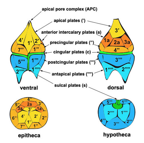
Fig. F. Kofoid system of tabulation. © Mona Hoppenrath
Although the Kofoid System is usually easy to apply, ambiguities in the attribution of some plates to one series or another can cause problems. This, combined with the mechanical, consecutive numbering, renders the system poor for intergeneric comparisons. Taylor (1980) introduced an alternative model of plate nomenclature (Figs will be added), elaborated on by Evitt (1985). It consists of three epithecal polar (A–C), six pre-equatorial (1–6), six equatorial (a–f), six postequatorial (I–VI), and three hypothecal polar (X–Z) sectors, which represent hypothetical primary plates from which homologous plates can be recognized by assuming subdivisions, suture losses, and plate size and position changes. The first step is to normalize the cell to a sphere, removing obvious plate distortions. Then the primary plates and their sutures are determined by studying the relationships of the plates to each other (see examples given by Evitt 1985).
The cingulum divides the cell into an anterior body portion, the episome (epicone, epitheca) and a posterior hyposome (hypocone, hypotheca). If the distal and proximal ends of the cingulum do not meet at an equal level at the sulcus they are said to be displaced. Displacement may be left-handed (the most common condition), in which the proximal (left) end is more anterior, or right-handed. The degree of displacement is measured in cingular-widths, given from the upper edges. The sulcus can extend on the episome and stops at the posterior of the cell. In athecate (wall-less<) cells there is a thin anterior groove, the acrobase, which reaches the cell’s apex. Acrobases can be straight, sigmoid, or form loops around the apex of the cell.
During cytokinesis the plane of cell cleavage is typically oblique. In thecate species the theca may be shared by the daughter cells, with synthesis of the missing components (desmoschisis), or the parent theca may be cast off, each daughter cell forming a complete new theca (eleutheroschisis).


