Drawings below are first made from photographs. The drawings should then be compared, under a microscope, with the preserved squid from which the photographs were made. This reduces the errors that can occur in interpreting the photophore distributions. Use caution, therefore, on this page in viewing the color and distribution of the dots (representing photophores) as their distributions have not yet been confirmed by comparing the drawings against the actual specimen under the microscope.

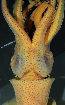
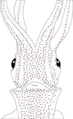

Figure. Ventral views of the integumental photophores. Left - Photograph of the preserved squid. Middle - Outline drawing from photograph with all integumental photophores represented by colored dots. Right - Photograph with colored dots superimposed on integumental photophores. Red dots - Complex photophores. Blue dots - Non-complex photophores. Images by R. Young.
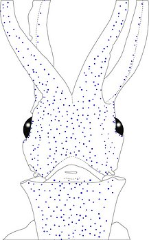
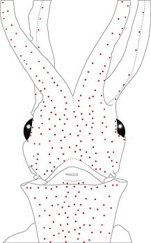
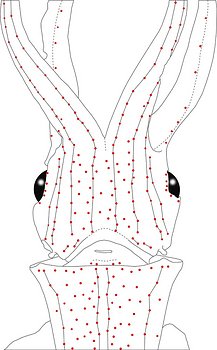
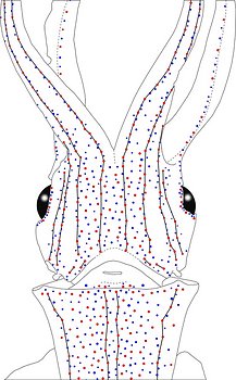
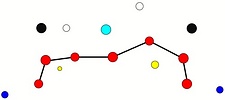
Figure. Same squid, same view as above. Left - Drawing showing only the non-complex (blue) photophores. Left middle - Drawing showing only the complex (red) photophores. Right middle - Same as previous drawing with lines connecting red photophores to aid comparisons of photophore patterns between species. Right - Drawing showing all photophores and lines showing red photophore patterns. Images by R. Young.
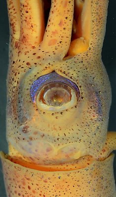
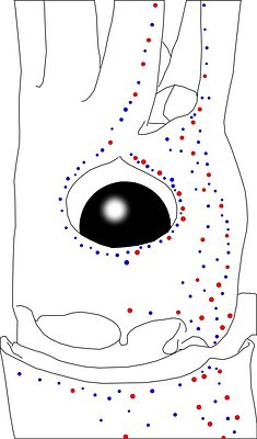
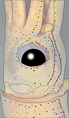
Figure. Side view of the head and proximal regions of the arms of the same squid (25 mm ML) as above. Left - Photograph of the preserved squid. Middle - Outline drawing from photograph with all integumental photophores represented by colored dots. Right - Outline drawing over dimmed photograph with all integumental photophores represented by colored dots, and with lines connecting some photophores. Red dots - Complex photophores. Blue dots - Non-complex photophores. Lines connecting photophores assist in understanding the organization of the photophores. Black lines - Patterns based on red photophores. Yellow line - Pattern based on red and blue photophores. Images by R. Young.
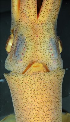
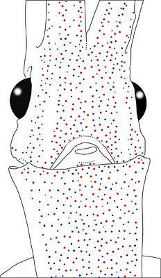

Figure. Ventral views of the integumental photophores . Left - Photograph of the preserved squid. Middle - Outline drawing from photograph with all integumental photophores represented by colored dots. Right - Photograph with colored dots superimposed on integumental photophores. Red dots - Complex photophores. Blue dots - Non-complex photophores. Images by R. Young.
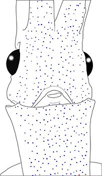
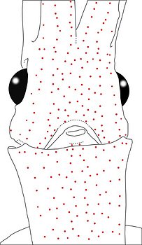
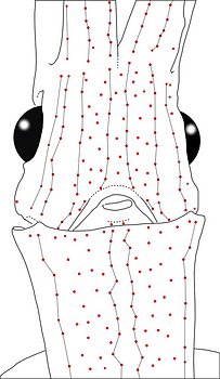
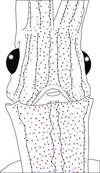
Figure. Same squid, same view as above. Left - Drawing showing only the non-complex (blue) photophores. Left middle - Drawing showing only the complex (red) photophores. Right middle - Same as previous drawing with lines connecting red photophores to aid comparisons of photophore patterns between species. Right - Drawing showing all photophores and lines showing red photophore patterns. Images by R. Young.
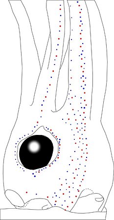
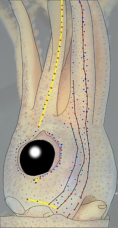
Figure. Oblique view of the head and proximal regions of the arms of the same squid (35 mm ML) as above. Left - Photograph of the preserved squid. Middle - Outline drawing from photograph with all integumental photophores represented by colored dots. Right - Outline drawing over dimmed photograph with all integumental photophores represented by colored dots, and with lines connecting some photophores. Red dots - Complex photophores. Blue dots - Non-complex photophores. Lines connecting photophores assist in understanding the organization of the photophores. Black lines - Patterns based on red photophores. Yellow line - Pattern based on red and blue photophores. Images by R. Young.
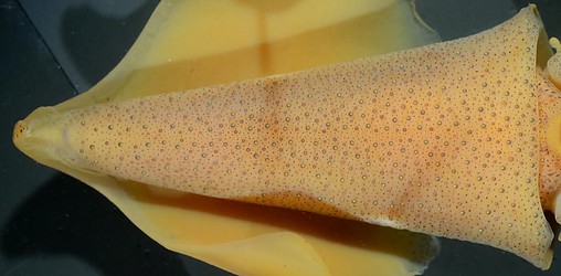
Figure. Ventral view of the mantle of the same 2 female squid as illustrated above. Top - 25 mm ML. Bottom - 35 mm ML. Note that the Median Mantle sector is fairly apparent posteriorly but not in the anterior 15-20 % of the ML.
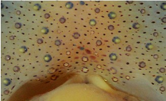
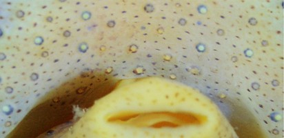
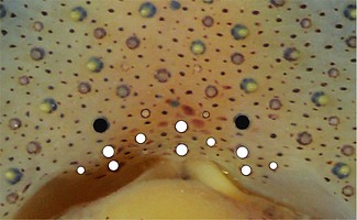
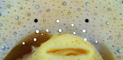
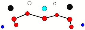
Table. Ventral view of the funnel-groove region of A. morisii. Top tier - Photograph of the preserved squid showing photophores of the funnel groove and surrounding tissue. Middle tier - In the photograph, white dots are placed over the "white" photophores of the funnel groove. Black dots are placed over the two most posteromedial "red" photophores of the ventral head to mark the anterolateral edges of the funnel groove. Bottom tier - Same pattern of dots as in photographs but dots are colored. Colored dots, lines - Aid in comparison of patterns between species. Open dots - Represent photophores that are either of uncertain status as "white" photophores or have variable presence. Images by R. Young. Comparison of white photophore patterns for all Abraliopsis species can be found here.
| Mature (?) female, 24 mm ML | Mature female, 39 mm ML |
|---|---|
 |  |
 |  |
 |  |
Comments: Summary of photophore distributions. (photophore definitions found here)
ARMS:
Medial Arm IV Series - Continuous; reaches 2/3 of arm length.
Central Arm IV Sector - Scattered blue photophores present near arm base.
Lateral Arm IV Series - Continuous; extends to arm tip.
Lateral Membrane Series of arm IV - Continuous; reaches at least 2/3 of arm length.
Arm III Series - Continuous; reaches at least 80% of arm length.
HEAD - Typical scattered pattern with:
Median Head Series - Absent.
Median Head Sector - Scattered red and blue photophores.
Medial Head Series - Arbitrary designation: About 7-8 red photophores at 25 & 35 mm ML.
Lateral Head Sector - Many scattered red photophores (9-11 at 25; 12-13 at 35 mm ML.
Lateral Head Series - Well defined.
Window Sector - Few scattered blues at either end. Otherwise without photophores.
1st Lateral Window Series - Continuous series with 8 red photophores at 25 mm ML, 10 at 35 mm ML.
2nd Lateral Window Series - Continuous series with 5 or 6 red photophores at 25 & 35 mm ML.
Eyelid Series - Red photophores only on ventral half of eyelid. Posterior Eyelid Series with one large blue photophores displaced slightly posteriorly from eyelid series. Formula: RbBB at 25 and 35 mm ML.
Occipital Series - RbRbR at 25 mm ML; RRbR at 35 mm ML.
Lateral Funnel-groove Series - Two nearly overlapping segments; each segment with 2 reds at 25 mm ML. At 35 mm ML, two overlapping segments; posterior segment with 4 reds, anterior segment with two reds.
Olfactory Photophore. Blue at 35 mm ML.
White Patch (funnel groove) - Moderate Pattern (red, green, blue + yellow with size).
FUNNEL: Patches not well demarcated.
Medial Patch - Scattered red photophores.
Lateral Patch - Two red photophores (in young at least - see photo on previous page).
MANTLE - Typical scattered pattern with:
Median Mantle Sector - Central bare strip poorly defined in anterior 15-20% of the ML: blue photophores obscure central bare strip.
Medial Mantle Series - Moderate to very irregular.
Mediolateral Mantle Sector - Scattered photophores; Lateral Mantle Series not apparent.
Mantle-angle Series - Barely recognizable.


